A Look at Imaging Tests
The medical field has impacted human health in innumerable ways, but perhaps none is more fascinating than imaging technology. Since the invention of the X-ray in 1895, these breakthroughs have allowed medical experts to see inside the body without surgery. Here is a breakdown of some of the more commonly used imaging tests that can help medical professionals identify internal issues.

X-RAY
The most widely used imaging device, an X-ray machine works by sending electromagnetic waves (low-dose radiation) into your body to detect things like broken bones and to see the internal structure of teeth. In addition, a dye may be used with X-rays to view blood vessels, a procedure called angiography. An X-ray is a quick test with quick results, but it also produces the most basic of medical images.
Computed Tomography (CT)
The deeper you delve into the body, the more precise the equipment and its imagery need to be, and a CT, or computed tomography, is an excellent example of this. Consider a CT scan (also known as a CAT scan) to be like an advanced, 3D version of an X-ray, as it provides cross-sectional views of organs such as the brain.
Ultrasound
Ultrasounds are commonly associated with pregnancies, but they are also used to view organs and detect blood clots. As its name indicates, this diagnostic tool uses high-frequency sound waves to form an image of the tissue or fetus being examined.
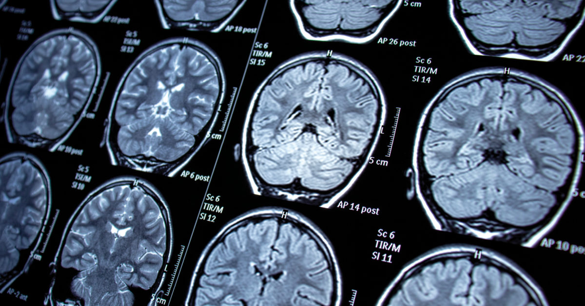
Magnetic Resonance Imaging (MRI)
An MRI produces detailed images of organs, bones, and soft tissue, but it does so through magnetic fields and radio waves. MRIs have a variety of diagnostic purposes but are most commonly known for detecting ligament tears in athletes. If you’re getting an MRI, be aware that you cannot have some metals inside or outside your body, the MRI tube is very confining, and the procedure can be quite loud during its fifteen to sixty minutes.
Be sure to ask your doctor any questions you may have about imaging devices and testing.